Difference Between 2D, 3D and 4D Ultrasounds
There are several types of ultrasound, or sonogram, procedures, but what they have in common is that all use high-frequency sound waves that scan and “bounce” off of the abdomen and pelvic cavity, resulting in an image of the fetus and surrounding placenta. According to the American Pregnancy Association, there are seven types of ultrasounds: transvaginal, standard (2 dimensional or 2D), advanced (targeting a specific issue of concern), fetal echocardiography (assessing the heart), Doppler, 3D and 4D (also known as “dynamic” 3D because it focuses on the face and fetal movements).
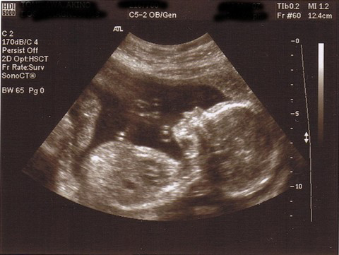 2D ultrasound of 20-week baby
Technically speaking, 3D and 4D ultrasounds are not “better†than 2D, just different. There are many diagnoses that can be made via 2D imaging that would not be possible with 3D and 4D views. However, when 2D and 3D or 2D and 4D are used in collaboration with one another, much information can be obtained.
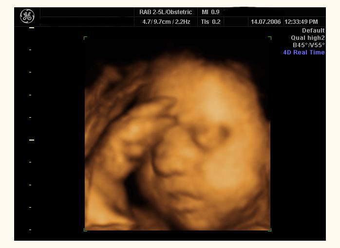 3D ultrasound
4D imaging was so named when it became possible to use ultrasound to obtain 3-Dimensional images in motion, as real time live-action videos. The 4th dimension is “time.†4D is able to capture two to four images per second, which is much quicker than 3D machines. Currently, General Electric is the only company to manufacturer a 4D ultrasound machine. So, if you’re visiting a lab or freestanding commercial fetal imaging site that offers 4D, they’re using GE’s Voluson 730 Expert.
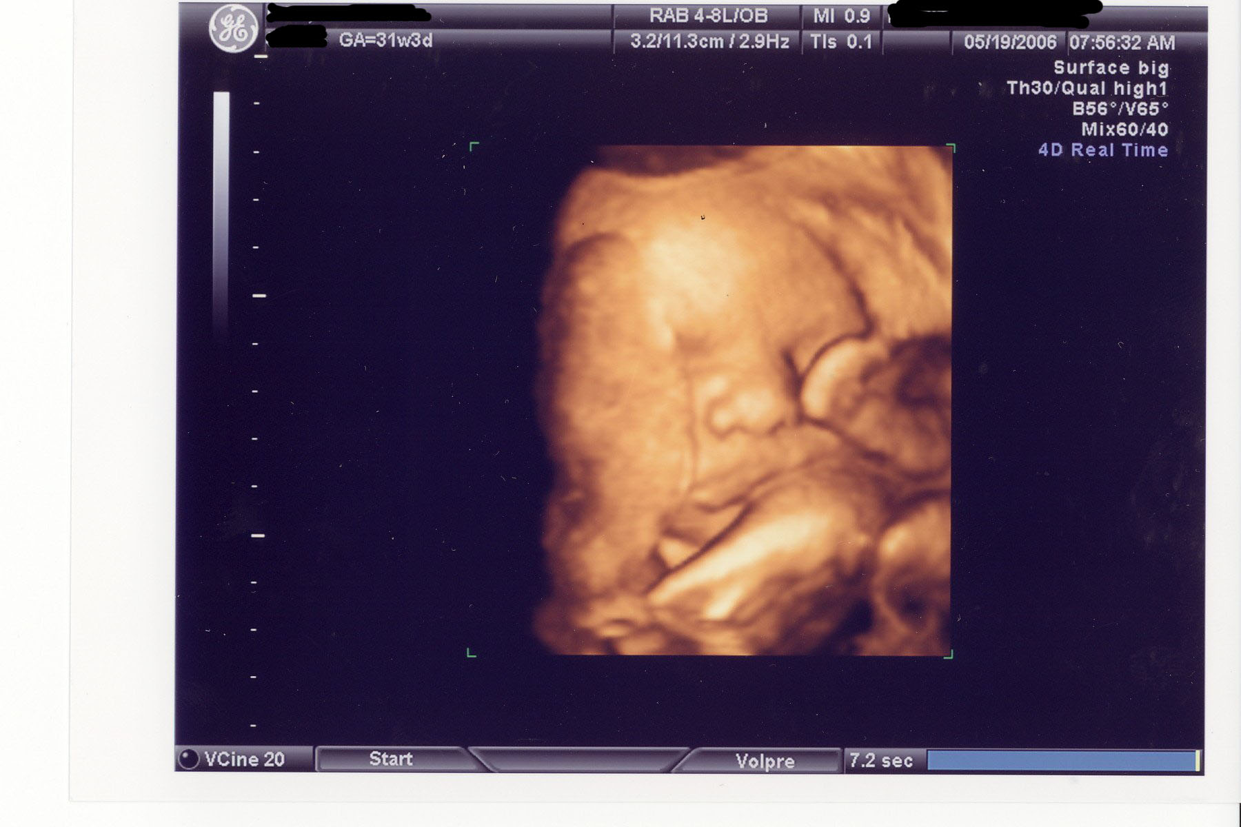 4D ultrasound of 31-week boy
For “reassurance” or “keepsake” ultrasound purposes, 4D is incredible in that it can offer a view of the unborn baby moving around, yawning, sucking his or her thumb and even appearing to “wave†a hand. When used in a diagnostic setting, 4D makes it possible to assess fetal development and/or diagnose issues in real-time, on organs that are moving before the sonographer or doctor’s eyes. 4D ultrasound can also be employed in improving accuracy during medical biopsies or amniocentesis.
[...] between 3D and 4D scans…after all, they look the same! Finally Googled it and found this explanation which clears things [...]
i am asking because a neice is 25 weeks and they orded a 4d and she has had a couple utrasounds and she has been very sick through this pregnancy (it is her first) she is borderline justational diabetic and she as well lost like 30 pounds in the first trimeater so ther are definately medical issues but im trying to understand the need for such an expensive procedure for one and the dr has not really said that there is a serious problem i was just wondering if you could tell me any general reasons for haveing them or could it simply be that the baby is extremely active and they cannot get the measurements that they need for the 2d ultrasounds
[...] any abnormalities. Â Â Â The first ultrasounds usually take place at the 18th to 20th week, the anomaly scan; it provides the parents with their first 2D images of the little one. Along with 2D images, [...]