What is 3D Baby Ultrasound?
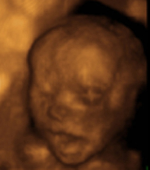
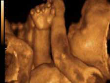
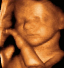
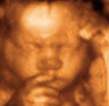
If you think traditional 2D black-and-white ultrasounds are neat, well, the 3D version is downright incredible when it comes to showing the appearance of a baby in the womb. By adding a third plane, the form of the baby takes shape and you can see facial features so well it’s possible to start pointing out exactly where he or she looks like Mom or Dad. Little feet and hands, too, are quite clear. Not to mention the “boy parts†and “girl partsâ€!
The advent of “live†3D ultrasound came in 1995, which, according to General Electric, sped up the time it took for an image to appear and show movement. The technology was further improved in 2000 and 2004 to make 3D ultrasound image-capturing even faster and more clear.
3D ultrasound involves using the same sound waves employed in 2D, but at different angles. This process, called “surface rendering,†allows software to immediately create a 3D image of the baby.
3D and 4D ultrasounds are not “better†than 2D, just different. There are many diagnoses that can be made via 2D imaging that would not be possible with 3D and 4D views. However, when 2D and 3D or 2D and 4D are used in collaboration with one another, much information can be obtained. For example, in 3D, conditions such as cleft palate and spina bifida are quite clear.
In recent years, freestanding fetal imaging centers have become quite popular. However, they vary greatly. Some employ experienced sonographers certified by American Registry of Diagnostic Medical Sonographers and perform diagnostic ultrasounds under a doctor’s orders, while also offering “reassurance†or “keepsake†ultrasounds commercially. Some just offer keepsake ultrasounds and may or may not forward the results to your doctor on request. The industry is not heavily regulated; to learn more about 3D ultrasound safety please continue reading Yourultrasound.com.