How Will Your Ultrasound Be Performed?
How your ultrasound is performed depends on several factors, including how far along your pregnancy is, where you’re having the ultrasound done, and if you’re having it for diagnostic or reassurance/ keepsake purposes.
As early as four and one-half weeks but more commonly at about eight weeks’ gestation, your obstetrician or midwife may do an ultrasound to confirm pregnancy and rule out ectopic or molar pregnancy. At this stage, it is often easier to observe fetal heartbeat and activity by performing a transvaginal ultrasound. This involves the insertion of a wand or probe, which most women don’t find uncomfortably large, into the vagina. The OB or technician will most likely apply a lubricant, or a lubricated condom-like sheath to make insertion more comfortable. By moving the transducer around, the OB can find the small, bean-like fetus in its gestational sac and identify the presence of cardiac activity, which presents as a “blink” of sorts. Measuring the fetus (“crown-rump length”) helps determine gestational age. If the heartbeat is visibly observed, general medical opinion is that the chances of the pregnancy continuing is greater than 95 percent. Most OBs keep a transvaginal ultrasound machine in their office, and can perform the ultrasound during a regular office visit. They can usually print this black-and-white image out as well, marking the area of cardiac activity with a “plus” sign.
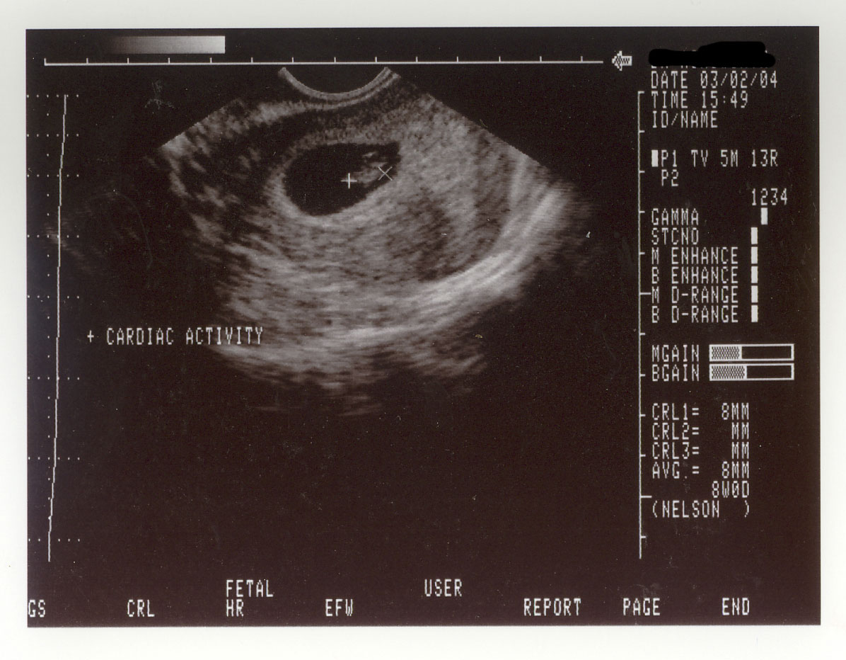 ultrasound at 8 weeks detects heartbeat
As early as 18 weeks’ gestation it’s likely that gender can be determined via ultrasound. If an ultrasound is medically indicated, or if your OB or midwife has fudged a bit so that you can get a look at the gender, he or she may send you to an outside facility, generally a contracted lab or imaging center, or a hospital’s radiology department. When you call to make your appointment they will tell you if you’ll be able to get a keepsake tape of DVD of the ultrasound, and they’ll advise you of one key point: Drink lots of water.
Water in the bladder pushes the uterus up, which makes it more accessible to the transabdominal transducer. All this water may make you need to go to the bathroom quite urgently, and the sonographer probably won’t let you, although some have been known to take pity and let you get up partway through the exam, once the major medical issue indicators have been checked. Seriously, you may have to pee so badly that you can’t even concentrate on the ultrasound monitor! Make sure you find out exactly how much water you need to drink and don’t go overboard. (Please note that if you’re interested in a non-medical, 3D or 4D reassurance or keepsake ultrasound, they usually don’t require that you drink any extra.)
Later in your pregnancy, if it is progressing normally, future visits often consist of the OB simply listening for a fetal heartbeat via a Doppler fetascope (also a form of ultrasound), checking the position of the fetus in the womb and perhaps assessing the level of amniotic fluid. If a transducer is used, expect to have gel applied to your abdomen to conduct the sound waves; the gel will probably be a little cold. Wear a two-piece outfit and you may not need to disrobe for the ultrasound.
The typical 3D or 4D exam takes 30 to 50 minutes, according to General Electric Healthcare. Various reassurance and keepsake ultrasound providers report that they take 30 to 60 minutes, depending on the “package†the customer wants and the positioning of the baby.